Leonardo Da Vinci Anatomical Drawings
LEONARDO DAVINCI ANATOMICAL FROM DRAWINGS THE ROYAL LIBRARY WINDSOR CASTLE THE METROPOLITAN MUSEUM OF ART NEW YORK
Views 134 Downloads 2 File size 18MB
Recommend stories


- Author / Uploaded
- michael
- Categories
- Leonardo Da Vinci
- Anatomía
- Perspectiva (Gráfica)
- Cráneo
- Dibujo
Citation preview
LEONARDO DAVINCI ANATOMICAL FROM
DRAWINGS
THE ROYAL LIBRARY WINDSOR CASTLE
THE METROPOLITAN
MUSEUM OF ART
NEW YORK
The exhibition has been made possible by a grant from Fiat S.p.A. and the Banca Nazionale del Lavoro, Rome, Italy. Additional support has been received from the N ational Endowment
for the Arts, Washington, D. C., a federal agency.
An indemnity has been granted by the Federal Council on the Arts and the Humanities.
Exhibitionheldat
THE METROPOLITAN
MUSEUM
oFART,NewYork
January 20, 1984, through April15, 1984
PUB LIS HE D BY
The Metropolitan Museum of Art, New York
Bradford D. Kelleher, Publisher John P. O'Neill, Editor in Chief Polly Cone, Editor Peter Oldenburg, Designer Copyright © 1983by The Metropolitan Museum of Art
Typeset by the Open Studio, Rhinebeck, New York. Printed and bound by Rae Publishing Co., Inc., Cedar Grove, N.J.
AHdrawings reproduced by gracious permission of Her Majesty Queen Elizabeth II ON THE COVER: Male nude seen from the back. Catalogue number 50
CONTENTS
F oR E W O R D
Philíppe de Montebello
5
PR EFA e E Robín Mackwotth- Young
6
Cario Pedretti
8
INTRODUCTION LEONARDO
DA VINCI THE ANATOMIST
KennethD. Keele
COLORPLATES CATALOGUE
10
15
OF DRAWINGS
25
The Internal Organs (1- 2)
27
Early Anatomical Studies (3-7)
31
Head and Brain (8- II)
47
The Alimentary and Reproductory Systems (12- 19)
57
Muscles and Skeleton (20- 33)
81
The Heart (34- 38)
123
Comparative Anatomy (39-42)
135
Human Proportions (43-46)
143
The Nude (47- 50)
151
CHRONOLOGICAL
TABLE
163
GLOSSARY
164
TABLE OF CONCORDANCE
165
BIBLIOGRAPHICAL
167
NOTE
FOREWORD
T
HE USE OF superlatives to describe Leonardo da Vinci, the most sensitive and relendessly curious of men, is entirely justified. Moreover, his consummate draftsmanship and sovereign intelIigence are often best preserved and seen in his works on paperoThe Royal Library at Windsor Castle, with its collection of six hundred drawings by Leonardo, constitutes the richest enclave of this master's works on paper. In the spring of 1981, the Metropolitan Museum, in collaboration with the J. Paul Getty Museum in Malibu, California, mounted an exhibition of fifty of Leonardo's nature studies, an unprecedented and indeed generous loan from the Royal Library at Windsor. Quod erat expectandum, the exhibition was an enormous success. We now delight in offering our visitors another exhibition of Leonardo's drawings, a superior selection of fifty anatomical studies, works that are widely acknowledged as among the fmest of Leonardo's creations on paper. Remarkable for their scientific exactness, these works are moving testimony not only to the artist's probing and fecund genius, but also to the most ennobling of life's miracles -the human anatomy. Weare most grateful to Her Majesty the Queen, who graciously agreed to lend this particular core of Leonardo's work. Forthe selection of the drawings, we should like to thank Sir Robin Mackworth- Young, Royal Librarian, Windsor Castle, and the Hon. Mrs. Roberts, Curator of the Print Room, Royal Library. With respect to the installation of the exhibition in New York, 1should like to acknowledge the respective roles of James Pilgrim, Deputy Director of the Metropolitan,Jacob Bean, Curator ofDrawings, Helen Mules, Assistant Curator ofDrawings, andJohn Buchanan, Registrar, for overseeing the myriad aspects entailed in mounting the exhibition. We are also grateful to Paul Williams, London, who was assisted by Hamish Muir, London, for their skillful installation designo PHILIPPE DE MONTEBELLO Director, The Metropolitan Museum of Art
5
PREFACE
OF
ALL THE MEN of genius who played a part in the Italian Renaissance, none is more remarkable than Leonardo da Vinci. Master of any discipline to which he set his hand-painting, sculpture, architecture, anatomical dissection, engineering, music-he exemplified the spirit of inquiry about nature to which the vast corpus of modern scientific knowledge owes its origino The impact of his genius has been preserved for us more directly than that of most of his contemporaries by his extraordinary talent for drawing. This he used for recording his thoughts, experiences, and discoveries much as a diarist or a scholar uses words, thus preserving for a later age intima te access to the very workings of his mind. An idea is set down as it emerges, perhaps filling a vacant space on an already crowded sheet. A few strokes of chalk, pen, or stylus suffice not OIUyto record sorne outer object-or sorne product of the imagination-but also to invest it with an inner energy, often of striking intensity. Sometimes he is processing a detail for a larger composition, sornetirnes simply recording knowledge. Words are not excluded, but are usually supplementary, appearing as comment by the side of sorne sketch. One such comment on an anatomical drawing explains his method. The use of drawing gives "knowledge that is impossible for ancient or modern writers [to convey] without an infmitely tedious example and confused prolixity of writing and time." Nowhere is this method better exemplified than in the anatornical series of drawings, whose interest is as much scientific as artistic. In the primitive conditions of the late fifteenth century, and with no medical training, this astonishing man acquired a knowledge of human anatomy far in advanee of the medical profession of his day. And the studies in which he recorded his fmdings bear comparison as works of art with his exquisite portrayals of the exterior of the human forrn and of horses, or with his dramatic representations of mountainous landscapes. On Leonardo's death the contents of his studio, which included several thousand drawings, passed to his favorite pupil, Francesco Melzi, whose handwriting may be seen on 19B in the present exhibition. On Melzi's death (about 1570) most of the collection was bought by the sculptor Pompeo Leoni, who rearranged the folios and bound them into volumes. Leoni, who was court sculptor to the king of Spain, took sorne of the volurnes to Madrid. After his death in 1609, one volume, containing examples of every field in which Leonardo worked, was acquired by the celebrated English collector Thornas Howard, Earl of Arundel, who brought it to England. While it belonged to him sorne of its drawings were engraved by Wenceslaus Hollar. Arundel had to leave the country during the Civil War, and it is uncertain whether or not he took the volurne with him. According to Count Galeazzo Arconati, who gave other Leonardo rnanuscripts to the Ambrosian Library in Milan, drawings concerning anatomy, nature, and color were "in the hands of the King of England" before 1640. This statement, and the fact that virtually all the surviving anatomical drawings by Leonardo are now in the Royal Collection, having formed part of the volurne bought by Arundel, suggests that the volurne did not leavethe
6
country, but was acquired by King Charles I. Others have surmised that it may not have reached the Royal Collection until after the restoration of King Charles II, to whom it could well have been sold or presented by Sir Peter Lely, one of the keenest collectors of drawings of his day. By whatever route it reached the Royal Collection, it is recorded as being in the possession of Queen Mary II in 1690, ayear after she and her husband ascended the English throne asjoint monarchs. This volume contained all the six hundred folios now at Windsor. During the three centuries that they remained within its covers, those executed in chalk suffered considerably from rubbing. To prevent further damage, most of the single-sided drawings were laid down on separate mounts in the nineteenth and early twentieth centuries. This technique could not however be applied to the anatomical series, most of whose folios bear drawings on both sides of the paper. The only solution was to rehouse them in new bindings, which exposed them to the same dangers as before. In recent years a new technique, which eliminates these dangers, has been devised. Each folio is encased within two thin panes of transparent plastic sheeting, and the resultant sandwich is inserted into a thick cardboard mount fumished with openings on both sides. Thus sheathed, a folio bearing a drawing on either side can not only be safely handled, but can also be placed on exhibition. Before being mounted in this way the folios were examined and photographed under ultraviolet light, sometimes with remarkable results (see 3,4, SA and B, 6A and B, 7). A small selection of these drawings was exhibited in Washington and Los Angeles in 1976. The present much larger exhibition, which comprises about a quarter of the anatomical series, was shown in London in 1977 and later in Florence, Hamburg, Mexico City, Adelaide, and Melboume. The entries for the catalogue were prepared by Kenneth Keele and Jane Roberts. The entire series of anatomical drawings has been published in facsimile by Harcourt Brace Jovanovich, with a defmitive catalogue by Kenneth Keele and CarIo Pedretti that includes a full transcription and translation into several modern languages of all the manuscript notes on the drawings.
ROBIN MACK WORTH- YOUNG Librarían, Wíndsor Castle
7
INTRODUCTION
T
HESE ANATOMICAL DRAWINGS by Leonardo da Vinci frorn the Queens collection at Windsor Castle have been selected and displayed as a synthesis of Leonardo's contributions to art and science in a field of endeavor that occupied him for a period of nearly thirty years, from about 1485 to 1510-15. The material is subdivided into nine groups according to a convenient subject-matter classification. This arrangement contributes to an even distribution of highlights in the visual impact produced by the beauty and precision of the drawings, but upon closer scrutiny it will soon become apparent that this material is best appreciated from the viewpoint of chronology, which gives the element of time perspective in the unfolding of Leonardo's thought. It is indeed this arrangement, or approach, that characterizes the facsimile edition of the total collection of Leonardo's anatomical studies at Windsor. It is quite appropriate, then, that this exhibition should open with an introductory section of two drawings only -one early and one late-showing an extraordinary progress in anatomical knowledge as Leonardo moves from traditional sources of learning, namely Mondino, to combine them with knowledge acquired through dissection, thus giving vividness and intensity to his vision of the human body as a machine. Much ofwhat has become famous ofLeonardo's involvement with technology-his extraordinary way of presenting the operating power of a machine-comes to be projected into these images of the human body, in which one may even detect the same pulsating effect that Vasari said was to be perceived in the throat of Mona Lisa. Leonardo's training as an artist in the Florentine studios of the late quattrocento coincides with an emerging concern for the representation of the human figure in action. With the Pollaiuolos it was not only the matter of assembling a vocabulary of gestures and attitudes, but also of introducing a new sense of vitality in the line of their drawings, the quick and lively notation of movement replacing the slow, careful defmition of form: a quality of line that appeals to Leonardo as early as 1473, when he first discovers how light affectsthe vision of rocks, and trees, and waters, and fields, as he draws alandscape of the Arnovalley that is a prefiguration of his later approach to natural forms in terms of structure and of a continuous flow of energy. Leonardo's first codification of this new approach to natural forms and to thehuman figure comes with his Vatican Sto Jerome, the unfmished painting that dates from the time of the Adoration of the Magi, about 1480. This is often mentioned as evidence of an anatomical knowledge based on dissection. It is indeed amazing how the muscles of the neck and shoulders should be so brilliantly displayed as to bear comparison with the most skillful analysis of the same muscles thirty years later, in the drawings of about 1510 shown in this exhibition (e.g., 27A and B). But the StoJerome is above all the first document of Leonardo's principle of representing the human figure "in context," as if architecturaHy conceived in ground plan and elevation so as to enhance its volumetric presence in relation to its setting. This explains the extraordinary vitality
8
of all the paintings he was to produce later, when, for instance, the grotto of the Virgin of the
Rocks was to be made so intimately related to the character of the figures it envelops as to be immediately felt as an integral part of the iconographic program and not to be taken simply as a decorative backdrop. Whenever, in fact, the human figure is presented as the nucleus of a natural setting, the equation between the figure and the generative forces of nature is implied. Hence the parallel that can be drawn between the celebrated drawing of a child in the womb and a human figure surrounded by an enveloping landscape, as in the Mona Lisa, in Leda, and in the Virgin and Child with Sto Anne. And since generation is also transformation, the link is soon established with the stereometric principles in the architecture of the High Renaissance, when Bramante would conceive of a building as the nucleus of an enveloping architectural setting. Leonardo and Bramante were friends. Little is known of the interaction of their ideas, the outcome of which, however, is that period of grandeur and monumentality in Italian art that is rightly viewed as a revival of the ideals of antiquity. It is an expression of intellectual clarity and power that reflects the imperial ambitions of popes and monarchs and the civic pride that was the basis of the socially reorganized Florentine Republic. The splendor and majesty of forms, both human and architectural, postulated by the artistic principles of Leonardo and Bramante were to lead inevitably to the academic art inherent in Raphael's response to those principles. And with this carne the anxiety of an age of reforms that Michelangelo was best to express with his concept of the human body in attitudes of struggle. CARLO PEDRETTI
9
LEONARDO
To
DA VINeI
THE ANATOMIST
SAY THA T LEONARDO DA VINCI was a unique genetic mutation is perhaps only to put into modern languageVasari's sixteenth-century verdict that "his genius was the gift of God." But this gift or mutation was expressed not only in his intellect, but in his physique also. Moreover, Leonardo's approach to the anatomy of the human body was significantly influenced by his own remarkable physical attributes. According to Vasari, he combined in himself exquisite sensory sensitivity with great physical strength and dexterity, if we may use this term for a man whose writing and drawings throughout his life were left handed. And it happens that his work in anatomy falls into two clear-cut periods of his life that correspond to those sensory and motor attributes of his nature. Leonardo's remarkable genetic endowments were derived from a peasant girl, Caterina, and a Florentine notary, Ser Piero da Vinci. Their bastard son, Leonardo, was born at Vinci on 15 April 1452. From avery early age he is said to have shown exceptional ability in geometry, music, and artistic expression. Ser Piero, noticing this, took his son's drawings to his friend Andrea del Verrocchio in Florence. Verrocchio was so struck by their quality that he accepted the promising young man straight away into his workshop. Verrocchio himself was well aware of the importance of perspective in art as well as the potential enrichment to art of representing the human body by a knowledge of anatomy. Like Leonardo he was an unlettered man, unimpeded by traditionallearning from personal observation and the practical application of his ideas. In a neighboring bottega the brothers Antonio and Piero del Pollaiuolo were similarly anatomically minded, using their knowledge as a basis for such pictures as the Martyrdom oJ Sto Sebastian (London, National Gallery). Such a background provided Leonardo with an ideal point of departure for his own painting of St.Jerome (Rome, Vatican Museum), in which the anatomy of the head and neck so dramatically portrays the agony of his soul. Perspective, however, was a less congenial subject to Verrocchio, who was daunted by its geometrical requirements. Not so Leonardo, who forged ahead in this field, basing his studies on the work of Leon Battista Alberti and Piero della Francesca. From experimental observations of objects through a vertical glass pane, or pariete, Leonardo came to appreciate the relation between perspective and the quantitative or measured observation of external bodies in their true proportions. This realization led him to apply himself to the objective representation of machines with such a degree of measurable accuracy that they have been reconstructed in recent years. It soon occurred to Leonardo that the same perspectival principle could be applied to extract "true knowledge" from the "universal machine of the earth." And what applied to the macrocosm of the earth applied also to that microcosm, the living body of man, which like the great "terrestrial machine" is "enclosed in the sphere of air." Thus Leonardo's early explorations into human anatomy focused on the nature of "experience," and in particular of perspectival experience. It is mainly in these early years, about 1490,
la
that we fmd his many diatribes against the "authorities." For instance: "Many will think that they can with reason blame me, alleging that my proofs are contrary to the authority of certain men held in great reverence by their unexperiencedjudgments, not considering that my works are the issue of simple and plain experience which is the true mistress" (Codex Atlanticus, f.r 19va). This was written in those very same years during which he was carrying out diligent and painstaking experiments on perspective as well as his first anatomical dissections. In this first period of Leonardo's anatomical studies, from about 1487 to 1493, it is interesting to observe how dissections of the sensory nervous system, particularly those parts concemed with vision, predominate. This assertion is only apparently contradicted by the fact that the fmest drawings of the skull that he ever made were drawn at this time. One notices, however, that the text alongside these early drawings of the skull (as, for example, 8A) is largely devoted to the location of the center of the senses and vision within the skull. Here the system of crossing lines is mainly devoted to demonstrating the site of "the confluence of all the senses,"that is, the senso commune in which he locates the soul. And in the lower drawing on the same page the optic nerves fmd clear and isolated representation. Again, on the verso of this folio (8B), alongside his marvelous exposure of the orbit and maxillary sinus, he writes: "The eye, the instrument of vision, is hidden in the cavity above .... The hole b [the optic foramen] is where the visual power passes to the senso commune." It was thus that he gave anatomical reality to his description of the eye as "the window of the soul." This preoccupation with the physiology of vision even in such an unlikely anatomical context betrays the fact that the majority of Leonardo's many studies of the eye and vision are not to be found in his so-called anatomical manuscripts, but are scattered about elsewhere. All of them, however, are áimed at a deeper understanding of the nature of the subjective side of "experience" as obtained from all the senses, not only the eye. In parallel with this he was attempting to analyze the nature of the objective observation of natural phenomena, such as the shape, size, and distance of objects, using the technique of perspectival proportions on the vertical glass pane. Through the combination of perspective and physiology of vision Leonardo hoped to understand how "the mind of the painter must of necessity be transformed into nature's mind in order to act as an interpreter between nature and art" (Treatise on Painting, f.24v). Thus did Leonardo bridge the chasm between science and arto After he felt that he had achieved an understanding of how "experience" could act as an interpreter between nature and art, Leonardo abandoned his anatomical investigations for nearly twenty years. During these years he was developing his science of the macrocosm of the world, which he called the "terrestrial machine." Finding simple linear perspective inadequate for solving distance problems, he extrapolated the principle of perspective to color and aerial perspective. He also formed the view that similar perspectival rules could be further extended in nature to the realm of what he called "the four powers of nature": movement, weight, force, and percussion, acting on the four elements of earth, air, fire, and water. AH power, because it did not occupy space, he saw as "spiritual" forms of energy, manifested in movement or change. Having performed innumerable experiments with pulleys, levers, mirrors, lenses, and particularly with water, he carne to the conclusion that these powers obeyed a perspectival or "pyramidal" form of action ("All the powers of nature have to be called pyramidal." Codex Atlanticus, f. 15Ir-a). J
11
At first Leonardo applied his rules of mechanics to the movements of man's body as a whole-to his center of gravity, to the actions of walking, running, and swimming. Many such studies are to be found in his Treatise on Painting. About 1495 Leonardo became friendly with the mathematician Luca Pacioli, who prop-
ounded to him the works of such dassical masters of mathematics as Eudid and Archimedes. This tumed Leonardo's interest even more strongly toward the conviction that geometry held the key to the interpretation of nature; and this induded the effects of the "four powers" within the body of man and of animals. The opportunity to pursue anatomy further seems to have occurred by chance during one of Leonardo's visits to the hospital of Santa Maria Nuova in Florence. He tells how an oId man a few hours before his death told me that he had passed a hundred years, and that he did not feelany bodily deficiency other than weakness. And thus while sitting on a bed in the hospital of Santa Maria Nuova in Florence, without any movement or sign of distress he passed away from this life. And 1 made an anatomy of him in order to see the cause of so sweet a death .... This anatomy 1 described very diligently and with great ease because of the absence of fat and humors which much impede knowledge of the parts (R.L. 19027V). In the performance of anatomical dissection Leonardo experienced the satisfaction of putting both his artistic and scientific principles into practice. Consumed as he was by curiosity and a passion for investigation, he was never one who believed in science for science'ssake. At the end of one of his paeans in praise of science he condudes abruptly with the verdict: "From it [science] is born creative action, which is of much more value." And among his anatomical notes he comments: "This generation deserves .unmeasured praises for the usefulness of the things they have invented for the use of man: and would deserve them even more if they had not invented noxious things like poisons and other similar things that destroy life or the mind" (R.L. 19°45 v) . Thus when Leonardo returned to anatomical investigation he put into practice all the scientific knowledge acquired since his earlier studies sorne twenty years before. This time the focus of his attention was centered on the movements in man's body. He sums up his outlook well on R.L. 1906oR, alongside a drawing of the maternal and fetal circulations: Why nature cannot give movement to animals without mechanical instruments is demonstrated by me in this book on the actions of movement made by nature in animals. For this reason 1 have composed the rules of the four powers of nature, without which nothing through her can give local motion to these animals; and how this movement engenders, and is engendered by, each of the other three powers. . .. We shall begin by stating that every insentient local movement is generated by a sentient mover,just as in a dock the counterpoise is raised up by man, who is its mover.
Again, right in the middle of sorne beautiful drawings of the anatomy of the hand, on 3 2B, Leonardo suddenly breaks out with the injunction: "Make the book on the elements and practice of mechanics precede the demonstration of the movement and force of man and other animals, by means of which you will be able to prove all your propositions." Leonardo's anatomical drawings can be looked at and enjoyed by many different kinds 12
of eye. For example, the eye of the artist will see in them the skill of his perspectival reduction of a three-dimensional object into a two-dimensional representation and the delicate hatchings of light and shade giving birth to the illusion of relief. The scientific eye will appreciate the three-dimensional accuracy of what he portrays: his insistence on the demonstration of all parts from at least three aspects, from in front, behind, and the side. To this he added his uncanny power of illustrating the mechanics of the movement of joints and muscles. Leonardo himself gave most diligent attention to developing his technique of anatomical illustration. One of many such passages (abbreviated) runs: This plan of mine of the human body will be unfolded to you just as though you had the natural man before you. The reason is that if you wish to know thoroughly the parts of man after he has been dissected you must either turn him, or your eye, so that you examine him from different aspects, from below, aboye, and from the sides .... But you must understand that such knowledge as this will not continually satisfy you on account of the very great confusion that must arise from the mixture of membranes with veins, arteries, nerves, tendons, muscles, bones, and the blood that itself tinges every part with the same color .... Therefore it becomes necessary to have severaldissections: you will need three in order to have a complete knowledge of the veins and arteries: three others for a knowledge of the membranes: three for the nerves, muscles, and ligaments; three for the bones and cartilages, and three for anatomy of the bones, for these have to be sawn through in order to show which are hollow and which not .... Three also must be devoted to the female body, and in this there is great mystery by reason of the womb and its fetus ... (R.L. 1906IR).
There is good reason to believe that Leonardo in fact carried out a great part, if not all, of this plan, for late in life he mentioned that he had dissected more than thirty bodies, and it is known that a number of small notebooks (libretti) offered to and refused by the grand duke Cosimo II de Medici in Florence in 1613 have since been lost. For particular anatomical problems Leonardo devised ingenious solutions. For example, in order to clarify the action of joints as fulera for movements of the bones, he separated their surfaces. "Thus you will give true knowledge of their shapes, knowledge that is impossible for either ancient or modem writers without an infmitely tedious example and confused prolixity of writing and time" (20A). When confronted with the problem of demonstrating the relations of muscles on different planes he advocates: "Before you form the muscles make in their place threads that should demonstrate the positions of these muscles; the ends of these [threads] should terminate at the center of the attachments of the muscles to the bones" (27B and R.L. 19°1 7R). Thus he reduced bones andjoints to levers acting on fulera, and muscles to lines of force acting on these levers. When wishing to reveal the true shape of the ventricles of the brain, Leonardo utilized his skill in sculpture, making wax casts of them by injection, and then removing the surrounding brain tissue (10). When wishing to observe the movements of blood as it streamed out through the aortic valve of the heart, he made a glass cast of the part (38). In summary, Leonardo's anatomical drawings were ofthree main types: those derived from his medieval predecessors, drawings of" descriptive anatomy" from untrammeled observation, and drawings illustrating his own physicallaws applied to the human body.
I3
Anatomy in medieval times was very crude. Primitive squatting figures containing almost unrecognizable organs meant to be read symbolical1y, not as true representations, were standard works used by doctors. Sorne of Leonardo's ear1y anatomical drawings of about 1489 were founded on this medieval anatomy. For example, the drawing of the three cerebral ventric1es (9A) represents them according to the pattern found in The Philosophy 01 Albettus Magnus, a copy
of which Leonardo possessed. From another medieval work, Ketham's Fasciculus 01 Medicine, Leonardo borrowed the outline of the human figure into which he inserted his own visualization of the "tree of the vessels" based upon the concept of Galen (1). In this same period he was making purely descriptive anatomical illustrations based on his own observations and made possible by his mastery of perspective. Such were those of the skull on R.L. 19°5 8v, where for the first time the maxillary sinus in the cheekbone is demonstrated. This kind of illustration, based on direct dissection, was quite unprecedented. By the eventual application of his physicallaws to human anatomy, Leonardo achieved a wholly unique penetration into the mechanical principles of physiology. His contemporaries were quite unready for this. Indeed, descriptive anatomy itself did not come to the surface until Galenic anatomy was undermined by the successful revolutionary efforts of Vesalius in his magnificently produced work, De Humani Corporis Fabrica, published in 1543, twenty-four years after Leonardo's death. This was the work for which contemporary anatomists were ready; this was the work that historically opened the floodgates of future anatomical progress. Thus in anatomy, as in so many other aspects of his life'swork, Leonardo was aman who awoke too early in the dawn of the scientific Renaissance while others still slept. Leonardo unconsciously described himself and his own particular genius for anatomy in the following passage (R.L. 19°7° V): You who say that it is better to look at an anatomical demonstration than to see these drawings would be right, if it were possible to observe all the details shown in these drawings in a single figure, in which with all your ability you will not see, nor acquire knowledge of more than a few vessels.... And as one single body did not suffice for sufficiently long a time it was necessary to proceed by stages with as many bodies as would render my knowledge complete: and this I repeated twice in order to discover the differences. But though possessed of an interest in the subject you may perhaps be deterred by natural repugnance, or if this does not restrain you then perhaps by the fear of passing the night hours in the company of these corpses, quartered and flayed, and horrible to behold. And if this does not deter you, then perhaps you may lack the skill in drawing essential for such representation, and even if you possess this skill it may not be combined with a knowledge of perspective, while if it is so combined you may not be versed in the methods of geometrical demonstration, or the methods of estimating the forces and power of the muscles; or you may perhaps be found wanting in patience so that you will not be diligent. Conceming which things, whether or no they have all been found in me, the hundred and twenty books which I have composed will give their verdict, yes or no. In these I have not been hindered either by avarice or negligence, but only by want of time. Farewel1. KENNETH
D. KEELE
COLORPLATES
8A. Two views of the skulI (R.L. 19057R)
17
8B. Two views of the skull dissectedto show the cavities of the orbit and maxillary sinus (R.L. 19057V)
18
1"
Ioff),," Q
..'"
...,:rrr-:~; Il~~l'ffh1 jI)
~"vf •
... ,.",
flf¡", ~1"1-
rst«
•
I,,-r..,c)r, "11" ~b('I'( --
(lM"'O
19A. The infant in the womb (R.L. 19I02R)
~ 1
20
27 A. The surjace muscles of (he neck and shoulder (R.L. 19003R)
A"'.' '.1
.
.. : "..~I.6t lIotI
r Mf :;
'!_ ",
1.~1~' VI') ¡~,jo1·rI"" "ullt{"',fltl
..J¡¡,
,r
1,
••] '"
j'
',1 tn', ;;!fl·.(·~j.}
" • tnll"rIi"\ JI
,,.JI "'1:+-
' ..~
27 B.
J!..
The muscles of the shoulder (R.L.
1900 3V)
21
47· Muscles
22
cif the arms and legs (R.L. 1263 7R)
48. Study of anude man lunging to the right (R.L. I2623R)
23
50. Male nude seenfrom
the back (R.L. 12596R)
CATALOGUE
OF DRAWINGS
Measurements are given in millimeters, height preceding width. The drawings are on white paper unless otherwise indicated, The catalogue has been arranged in roughly chronological sections, except for the last three, which act as an appendix to the theme ofLeonardo's anatomical studies.
THE INTERNAL
ORGANS
A
PERIOD of about ten years separates the two drawings shown here (r and 2). During these decades, which span the turn of the fifteenth century, Leonardo bridged the gap between medieval and modern anatomy. But he did not achieve this feat all at once. The advances made can be seen here in the greater accuracy in the location and shape of the liver, kidneys, and spleen demonstrated in the later drawing. The rather unhappy-looking man drawn in 1,in which Leonardo has retained a considerable amount of medieval anatomy, is taken from a medieval fasciculus of medicine.
27
1. Anatomical figure showing heart, lungs, and main arteries Pen and dark brown ink with colored washes over black chalk. 280 x 198 mm (R.L. 12597R)
2.
Dissection of the principal organs and arterial sy stem of a woman Pen and ink wash over black chalk on color-washed paper. Pricked through. 478 x 333 mm (R.L. 12281R)
This sheet is headed, between the legs of the figure: albero dj uene (tree of the vessels), and to the left,parte spiritualj (spiritual parts), relating to the Galenical vascular system, according to which the natural spirits were carried by veins from the liver and vital spirits by arteries from the heart. Below this is a reminder to "cut through the middle of the heart, liver, and lung and the kidneys so that you can represent the vascular tree in its entirety." The degree of anatomical inexactitude together with the ornamental handwriting suggest a date in the 1490S for this drawing, which relates to the ancient and medieval anatomical knowledge of Leonardo's predecessors. It is possible that it is connected to an anatomical model, for it is close in date to 16B, on which Leonardo makes a reference to the construction of such a model: "When you have finished making the man, you will make the statue with all its superficial measurements."
This study dates from sorne ten years later than l. While showing much progress in correcting sorne of the inaccuracies of the earlier drawing, it retains many of the errors Leonardo inherited from Galen and Avicenna. An example of this is the depiction of the vena cava passing the heart and giving a branch to the right ventricle, with no atrium at all , Original observation is shown in the relative accuracy of shape and size of the kidneys, liver, and spleen, which were studied in detail in the pages of Folio B. In the manuscript passage in the top left-hand comer Leonardo makes one of his many notes on further drawings to be done from his dissections: "Make this demonstration also seen from the side, that knowledge may be given how much one part may be behind the other, and then make one from behind, that knowledge may be given of the veins possessed by the spine, and by the heart, and greater veins." The manner of pricking through and folding of this sheet suggest that the main shape of the left-hand side of the page was drawn on first, the sheet folded vertically and pricked through, and that the right-hand side was drawn on the basis of these pinpricks. When the figure had been completed, and the asymmetrical features added, the whole figure was agam pinpricked, this time for transfer to another sheet (R.L. 12280).
I P. 'J.I't. ftt )";. f'
---
-,
«'fA •
1
•...
.,."1
,,.-4 '".ota, "'t'l 1'" .lrl't4
.1
,;
,IQ!.,., _ -,r;J ·.b·~
• Ih'. JJP'1·,o1·,I.tl..... I.¡ftN , -
_- . Aj¡il"llf('
'trI
4I~I~
,
Jo
rr.(K Jo
.. \
"•.,'11 ...."...... .. • "O,,!
rn" 'J
&
'I"',I~
>1"'1" ..l·'''.r
,",,1
n~l..,tI_.(':'~"" ,-,
~"..
t .. """"
.ift
........
J4,,yfoll'll,.,,·
niI'''r4
~",l, ..... ft'f
.,..I"JII''')#'.
..
,..
;lI\ lIIfp,
II'I',."·l~ ~..... IA-f""
2
EARLY ANATOMICAL
THIS
STUDIES
SECTIONconsists of five sheets of metalpoint studies on blue prepared paper, sorne of which (e.g., SA) are labeled with double letters in a sixteenth-century hand. They clearly date from the same chronological period (about 1487- 89), and the drawings on them are closely similar in contento The studies are mainly anatomical and show a combination of traditional views (particularly with reference to the brain) and the artist's own experimental anatomy, involving dissection in one caseof a frog (SE), in another of a monkey (SA), and in others of aman. These drawings are sometimes accompanied by Leonardo's records of his anatomical methods, in which he mentions what he has already done and what he has still to do. He is concerned in particular with problems regarding the nervous system. Our reading of these sheets has been greatly assisted by their examination under ultraviolet light. Whatever the substance of the metalpoint Leonardo used (it has always in the past been called silver), it must have faded from normal vision very soon after these drawings were made. Confirrnation of this is found in Dürer's copies of drawings on SE in his Dresden sketchbook, in which he drew only as much as is visible today, and not the complete metalpoint drawings, which are visible only under ultraviolet light.
3
Miscellaneous anatomical drawings Pen and dark brown ink (two shades) over metalpoint on blue prepared papeL 222 x 290 mm (R.L. I2627R)
32
The drawing bottom center shows Leonardo's attempts to acquaint himself with the general positions of the main viscera (labeled with indicator lines) in the chest and abdomen - what was known as a "situs figure" in his day. To the left of this, in an oval section, is an attempt at cerebrallocalization in the ventrides of the brain; the optic, olfactory, and acoustic nerves being traced back to their hypothetical destinations. Under ultraviolet light the ventricles of this diagram can be seen to be marked, from top to bottom: memoria, chomune senso (i.e., senso commune), and imprensiva. The three ventricles are drawn again within the skull in the old man's head to the left. Another drawing of the skull, apparently connected with the old man's head on the left margin, is clarified under ultraviolet light immediately to the left of the situs figure. It shows the orbits from which the optic nerves issue, converging on the anterior ventricle. This is connected by a narrow channel to the middle and posterior ventricles. For the diagrammatic treatment of the cerebral ventricles, seeentry for 9A, of I493-94. To the right, upside down, is a dissection of the leg, showing the saphenous nerve and its accompanying vein, with the straplike sartorius muscle labeled lacerto. The rough sketch in the center represents visceral organs of an animal. It
(liver), and sangue (blood), the latter indicating what appear to be two pleural cavities containing blood. Along the top of the sheet is a sketch of the lumbar spine, pelvis, femur, and tibia, drawn to show the relation of the bones to the origins and course of the sciatic nerve. The femur with its trochanter tertius, laterally, shows that Leonardo had sorne knowledge of the skeleton of the horse at this time. Several other totally new drawings appear under ultraviolet light. In the top right-hand comer are two sketches for a system of gear wheels and pinions for augmenting lifting power. To the left of these is Leonardo's familiar doodledjmmj (tell me), and below there is a sketch of nerves emerging from beneath the clavicle into the arm (compare 5A). To the right of the situs figure, bottom right, is a small interlacing pattern. Beneath the old man's head on the left-hand margin ís a rectangular structure: perhaps the ground plan of sorne architectural project. Below the sketch of the víscera in the center, facing toward the top of the sheet, a youthful profile has appeared. In addition, four enigmatic lines of script are revealed (top left), presented in the form of a riddle which has not yet been solved by Leonardo scholars. is labeled, from right to left, stomacho (stomach),fegato
3
ULTRAVIOLET
33
4. Muscles, tendons, and veins of the arms and legs Pen and brown ink (two shades) over two types of metalpoint on blue prepared paper. 2I2 x 297 mm (R.L. I26I7R)
The sheet contains eight drawings of a man's leg, including one segmental view (to left of center) and below this a sectional view of the lower leg, lettereda-o. To left and right, on the lower part of the sheet (turn the paper 90 degrees), are two studies of the arm and shoulder, showing veryelementary patterns of the brachial plexus. To the right of center is a rough sketch of the abdominal muscles, with ribs and pelvis. AH these forms are greatly clarified by ultraviolet light, as are the architectural plans along the lower edge of the sheet. Their purpose is still unknown, but they may refer to plans of palaces as shown in Codex Atlanticus, f.324r of about 1482, on which Leonardo listed the works that he was to take to Milan or leave in Florence. As one ground plan hints at columns and steps as in an antique temple, these architectural scribbles may be theoretical notations inspired by the reading of Vitruvius.
/
34
35
5 A. The bones and tendons
of the arm and shoulder with studies of animal vtscera Pen and dark brown ink over metalpoint on blue prepared paper. 7.22 x 304 mm (R.L. I2613R)
The drawings on this sheet are closely connected with those of 3: the views of animal viscera occupying most of the left-hand side of the sheet, and the faint studies of shoulders and the inverted leg at the right-hand edge are very similar to forms on that sheet. Once again, ultraviolet light has revealed a large number of these forms and clarifies various passages of the manuscript. That in the top right-hand comer, written from left to right, reads as follows: Jn questo modo nascho no i nervi dj tutta la persona a ciaschu nodo de la schiena
(In this manner originate the nerves of the whole person at each projection of the spine.) On the same subject a drawing of vertebrae, center lefi, invisible under ordinary light, is inscribed: osso / spuga / nervo voto (bone, sponge, empty nerve). This figure makes clear that the spinal nerve issues between two transverse processes of neighboring vertebrae. The term "empty nerve" refers to the concept of nerve impulses passing down a hollow ("empty") cavityalong the length of the nerve .
•
5A U L TRAVIOLET
37
5B. The skeleton and tendons of the arm and leg Pen and dark brown ink over metalpoint on blue prepared paper. (R.L. I2613V)
The main drawings covering the right-hand side of this sheet were copied (in reverse) by Dürer in his Dresden sketchbook. For various reasons it is unlike1ythat Dürer saw this sheet, and it is more likely that he had accessto a copy (perhaps an engraving) after it. Whatever the case, he copied OIUy as much of the drawings as are visible today: the incomplete drawing of the skeleton and tendons of the leg is incomplete in Díirer's copy, while ultraviolet light reveals that Leonardo had completed it in his metalpoint underdrawing. It seems therefore that the underdrawing had already faded from normal vision in Leonardo's lifetime. Other drawings that appear under ultraviolet light include a tower-like structure to left of center, comparable to one in the Codex Atlanticus.
)........ __ ...
\
(
--
-\(
--......_
-
---
The drawing at left refers to the spinal cord of a frog. The annotations surrounding the drawing, written both left to right and right to left, include passages such as the following: "The frog instantly dies when its spinal cord is perforated. And formerly it lived without head, without heart, or any entrails or intestines or skin. It thus seems that here lies the fundamentum of motion and of life." Such words presuppose a program of study and experimento The spinal cord is labeled "generative power," referring to the belief held by Hippocrates and other later authorities (including Avicenna) that "the most active and thickest part" of the semen comes from the spinal cord (see also 16A).
5B ULTRAVIOLET
39
6A. The skeleton, muscles, veins, and cartilage of (he head and neck
Ultraviolet light reveals that not only the main figure on this sheet, but also the subsidiary one, middle right, are concerned in detail with the anatomy of the neck. The quickly sketched figure, bottom left, can now be identified as a diagram of the abdomen, and at the right of this form, along the lower edge of the sheet, are a grotesque head and a crouching figure.
Pen and brown ink, heightened with white, over two types of metalpoint on blue prepared paper. 202 x 287 mm (R.L. I2609R)
\.
I
••
6A ULTRAVIOLET (
.
'
(~./
(
\
~
\
•
~
f.,
. ·i
41
6B. View of a church
There are no anatomical drawings on this side of the sheet, but the architectural forms-so well clarified by ultraviolet light-are contemporary with the anatomical drawings of the series. The main drawing appears to show a cross section of a model in which three of the arms of a centrally planned building, topped by a cupola, are shown, with conches, triforium gallery, and paneled dado on apsidal ground plans. The drawing is almost certainly related to the Romanesque church of San Sepolcro in Milan, which is referred to in other Leonardo manuscripts of this time. It is possible that the subsidiary sketch to left of center, revealed by ultraviolet light, indicates the actual structure of San Sepolcro, while the main study incorporates a restoration program inspired by Bramante .
Pen and dark brown ínk over metalpoint on blue prepared papeL (R.L. 12609V)
•
I
"
I
6B ULTRAVIOLET
43
7.
The musculature of the leg and the anatomy of the head and neck Pen and brown ink (two shades) and metalpoint on blue prepared paper. 203 X 300 mm (R.L. I2626R)
) \
The right-hand figure of the thigh and upper leg is annotated as follows: "1 have removed the muscle a-n (sartorius), which has a length of half a braccío, and 1have uncovered r-t. Now attend to what remains below m-o." The drawings of the skull and neck continue Leonardo's inquiries seen in 6A and repeat the traditional figure of the cerebral ventricles as seen in 3 (and in 9A). The labeling of the ventricles is altered in 7. From top to bottom it is: memoria (memory), senso commune and volota (senso commune and willpower), impresiva and inteletto (feeling of impressions and intellect). The architectural sketch, bottom left, appears to be related to Battagio's Santa Maria della Croce at Crema, of about 1493. The main building is apparently placed on the shore of a lake and is connected by a bridge to a round platform on a promontory on the opposite shore.
Ir
I
,
\ 1~ .
)
I
\
't'
\
44
\
..........lH'f)~l.j...,.-
1 -'
~
(
\
'~Cl1
7 ULTRAVIOLET
45
HEAD AND BRAIN
LEONARDO'S first fully worked anatornical studies were made on the head, about 1489. In these his prime objective appears to have been the location of thesenso commune, the meeting place of all the senses and the seat of the soul, which is located on 8A in the region of the third ventricle of the brain. In the course of his dissections of the skull and brain, Leonardo made severa! discoveries, unknown before his time, such as the maxillary antrum (8B) and the frontal sinus (8A and 9A), and he illustrated the optic chiasma (I I). The superb drawings of the cranium made in 1489 (e.g., 8A and B) are apparently the only detailed studies of the skull made by Leonardo. He evidently considered that he had mastered the correct anatomical depiction of this area.
47
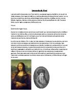
8A. Two views of the skull Pen and brown ink (two shades) over black chalk. 188 x 134 mm (R.L. 19057R)
8B.
Two views of the skull dissected to show the cavities Of the orbit and maxillary sinus Pen and brown ink over black chalk. (R.L. 19057v)
In these drawings of the human skull Leonardo exerted all his artistry in the perspectival creation of verisimilitude, as shown by the fine delicacy with which he represents variations in the contour of the skull bones in the upper drawing. The skull is shown from the side with the vault bisected in a medial sagittal section. In the lower figure the skull has been tilted forward and the section has been carried through the whole skull, so exposing the frontal and sphenoidal sinuses, the three cranial fossae, and the nasal cavity. It ends with an imaginative representation of the upper vertebrae. The squares with crossing lines dividing up the lower figure are best described by Leonardo's own words to the left of the sheet: "Where the line a-m is intersected by the line c-b there the meeting place of all the senses [senso commune]is made, and where the liner-n is intersected by the lineh-f there the fulcrum of the cranium is located, at one third from the base line of the head." In another place Leonardo states that "the senso commune is the seat of the soul," and we may therefore see in this figure his exact location of the soul in the body. The drawings can be dated 1489 owing to the close relationship between content and style of this sheet and that of another sheet of drawings at Windsor (R.L. 19059R) inscribed with the date 2 April 1489. The upper figure of the skull was engraved by Wenceslaus Hollar in 1645, when the drawing was in the collection of the Earl of Arundel.
Below the upper drawing Leonardo writes: "1wish to lift off that part of the bone armor of the cheek, which is found between the four lines a-b-c-d and through the exposed opening to demonstrate the width and depth of the two empty cavities that are concealed behind that [bone]." This exposure, illustrated in the lower drawing, shows the bony cavity of the orbit (marked d) "in which the instrument of vision is hidden." The cavity below this is the maxillary antrum or sinus (marked m) which, as Leonardo puts it, "contains the humor that nourished the roots of the teeth." Until these drawings were first thoroughly studied in 1901 it was believed that the maxillary antrum had been discovered by the Englishman N athaniel Highmore in 1651. In the notes below the lower drawing Leonardo records the position and nature of the vessels entering the two cavities. The lower cavity "receives veins into it through the holes m that descend from the brain passing through the sieve [cribriform plate of the ethmoid] that discharges the superfluous humors of the head into the nose. No other holes are evident in the cavity aboye which surrounds the eye. The hole b [optic foramen] is where the visual power passes to the senso commune; the hole n is the place from which the tears rise up from the heart to the eye, passing through the canal of the nose." This last passage is explicable by Leonardo's current belief that heat raised all fluids to the head.
8A
49
8B
5°
9A. The layers of the scalp compared with an onion Pen and brown ink (two shades) and red chalk. 203 x 1'52 mm (R.L. I2603R)
9B. Perspective studies Of the head Pen and brown ink with oxidized white and traces of metalpoint (R.L. I2603V)
These early drawings are well described in Leonardo's own words along the left-hand edge: "If you will cut an onion through the middle you will be able to see and count all the cover or rinds which circularly dothed the center of this onion. Similarly, if you will cut through the head of a man in the middle , you will first cut the hairs, then the coating and the muscular flesh and the pericranium, then the cranium, and within, the dura mater, and the pía mater and the cerebrum, then again the pia and the dura mater and the rete mirabile and the fundamentum of that, the bone," These layers are labeled in the main figure and in the section bottom right. Above the eye the frontal sinus is seen-which, with the superciliary arch, is given rather undue prominence by the artist (as in 8A and B). The frontal sinus was one of Leonardo's many anatomical discoveries. From the similarly "onion-coated" eyeball the optic nerve runs to the cavity representing the anterior of the cerebral ventrides. Bottom right is a horizontal sectional diagram of the upper part of the skull, with the top part hinged backward. The shape and form of the ventrides in these figures are diagrammatic representations of the description of the brain given by the old anatomists. Leonardo reached a much closer approximation to their actual form in 10, over a decade later. The date of the drawings on the present sheet is about 1493 - 94.
It has often been claimed that the group of drawings on the lower half of this page represent studies of spectacles. In fact they are studies in perspective of the optic nerve and auditory nerves converging within the brain onto the senso commune and are therefore related to the drawings of the cerebral ventricles on the recto of the sheet. At this time-that is in the years after 149o-Leonardo was attracted by the relationship between perspective, vision, and objective observation. The three miniature heads (bottom right) show an experiment in "transformation" whereby the projection of an anterior view can be used to construct views in perpendicular planes. This page contains a number of metalpoint studies that are only legible when viewed under ultraviolet light. The inverted profile at bottom, right of center, has been gone over (not by Leonardo) in white lead, which has since oxidized.
53
10. The brain injected to demanstrate the shape af the cerebral ventricles Pen and brown ink (two shades) with black chalk. 200 x 262 mm (R.L. 19127R)
11. The optic chiasma and cranial nerves Pen and brown ink (three shades) over traces of black chalk. 190 x 136 mm (R.L. 19052R)
The injection of parts to discover their true form is among the most important innovations in anatomical technique contributed by Leonardo. Here he applies it to the anatomy of the cerebral ventrieles. In the drawing top left the site of injection of melted wax is marked at the base of the third ventriele (here central, and labeled senso commune). The shape of the wax cast is seen in this and in the drawing to its right of the brain in horizontal section. It can be seen that the wax distorted the third ventriele and failed to fill the lateral ventrieles completely, but it revealed the first resemblance to their true size, shape, and position and represents a marked advance on the diagrammatic layout of the ventrieles in, e.g., 9A. The site of injection is again marked at the center of the rete mirabile in the bottom figure (identifying the brain as that of an ox) . The notes consist of detailed instructions of the technique of injection, which is again illustrated in the small sketch in the bottom right-hand comer. The drawings on this sheet are datable about I 508- 9.
The drawing top left shows the eyeballs, optic nerves, and optic chiasma. Parallel to and above the optic nerves are the olfactory nerves, about which Leonardo makes the following brief statement at the top of the page: "a b c d sono li neruj che pigliano liodori" (a bcd are the nerves which take up [and so, record] odors). In the main drawing to the right these features are placed in relation to the base of the skull and sorne of the cranial nerves. Leonardo showed great interest in the physiology of sensation, particularly visiono The main point of these drawings was to demonstrate the path of vision from the back of the eyeball to the base of the brain. This he achieved with remarkable success. The olfactory bulbs and nerves are also well shown, lying above the optic nerves. He demonstrates with praiseworthy accuracy the delicate motor nerves to the mus eles of the orbit, and the main (trigeminal) nerve to the face. The main body of manuscript on this sheet recounts the method of dissection that enabled the artist to make these drawings: Ease away the brain substance from the borders of the dura mater [the hardest of the three membranes surrounding the brain] .... Then note all the places where the dura mater penetrates the basilar bone with nerves ensheathed in it, together with the pia mater [the innermost of the three membranes surrounding the brain]. And you will acquire such knowledge with certainty when you diligently raise the pia mater, little by little, commencing from the edges and noting bit by bit, the situation of the aforesaid perforations, commencing first from the right or left side, and drawing this in its entirety; then you will follow the opposite side which will give you knowledge as to whether the previous one was correctly situated or noto Furthermore, you will get to understand whether the right side is the same as the left, and if you fmd differences, review the other anatorniesto see whether such a variation is universal in all men and women.
Leonardo was probably the first to draw the optic chiasma, in another sheet at Windsor datable about I500 (R.L. I2602R). The present sheet is datable about I 506- 8. In the lower drawing Leonardo is concerned with the blood supply of the uterus from the hypogastric artery, the remnant of which is shown curling upward toward the umbilicus.
54
_,
...,.
...., '
'1", -rr+
....".,..C'NHJ... :...,,·.II'T"'¡I~j ,,"
f"""""". hIMA" ',1'\
',Jo;. "(
.,.6.. .
c(rl,., '. .r.: ..1tI1) ·... {nlo "',r "". ('',A' """'A·"¡. ''''1 ' •
,.....1'1 ",..,.
,H"'/R'"
....
I«.,'~""
'1"""'''''' ..... l'"" "'' ' ,,', 1 ~" hf'1"f r« .),.",'" .... ,(' .. ~rf·,f ~/7)"" "1" ..... _ .. b.. .IrJt~
' t _J
" ..
"(~II
fU'ni' n II'r~
~" ••, h{ ,,1 'HfH1,.,tra .#fr.
"fu v"
~~O""".'.J- · ·rf 'rI) ~ ':al
_... ,"N,
f
..
,.
~~'T.~!' , t'U¡
¡..rl.. ~
, -
2IB
f¡" ,.., "doH
~ "lrJ
• t. ,,."..
J
,r'·' tr
I .~
22A. The brachial plexus and discussion concerning muscles Pen and brown ink (two shades) over traces of black chalk. 192 X 143 mm (R.L. 19020V)
22B. The mesentery Of the bowel and its blood supply Pen and brown ink (two shades) over traces of black chalk. (R.L. 19020R)
88
Two themes are considered on this sheet by Leonardo: the brachial plexus, described in the visual language of the drawing across the center of the sheet and the force of muscles, discussed in the notes. Since the study of the plexus on 2IA and B a little time before, Leonardo has evidently carried out further dissections, for the plexus is now correctly endowed with five (not four) roots, uniting to form upper, middle, and lower trunks. The main figure on this sheet is labeled del vechio (of the old man). On another sheet Leonardo gives details of how the old man died in the Hospital of S. Maria Nuova in Florence, and how he "made an anatorny" of his body (see page 12). There is good reason to date this event and the drawings of his dissected body (e.g., 22A) to 1508. The smaller drawings bottom right relate to the anterior vertebral muscles, labeled a bcd e in the main diagram and seen in cross section in the bottom right-hand comer. Just above the main drawing of the plexus Leonardo writes: "Any one of the five branches saved from a sword-cut is enough for sensation of the arm ," suggesting that the artist had clinical experience of such an event, perhaps gained during Cesare Borgia's campaigns of 1502- 3, in which Leonardo saw active service. The manuscript notes hardly concem the drawings at all and are instead devoted to a discussion dellaforza de mvsscholj (on the force of muscles), according to the heading. In his notes Leonardo foreshadows the more advanced study of musculature carried out by him a few years later in the pages of Manuscript A. At this stage he is intrigued by the fact that nerves, muscles, and arteries are easily broken, but at the same time capable of exerting considerable force. He explains this phenomenon by the presence of membranes surrounding the muscle fiber.
The notes and drawings on this sheet refcr to the Galenical system (adhered to also by Mondino and Leonardo) whereby chyle absorbed from food in the intestine is carried by the superior mesenteric and portal vein (seen in all but one of the diagrams) to the liver, where it is turned into blood; at the same time this vein "nourishes" the mesentery, beautifully drawn by Leonardo in the two central diagrams. The passage at the top of the page is one of several on miscellaneous pages of notes by Leonardo referring to his interest in fashion: "Long nails among Europeans are deemed shameful. Among Indians they are held in great veneration; and they have them painted with penetrating fluids and adorn them with various patterns; and they say that such practices are for gentlefolk and that short nails are for laborers and mechanics in different trades."
22A
•';"¡.I'
tf ",.. ... (1.'1 1"1",
~n
"'"
ftt·,,,
.
"''(',,,4,,,·, .... n¡)11
...n',)I, . ,i
'"}.L··h .....le" 22B
9°
~"',rrl
111\ ~1I
".J,
0)'--1'" , ..
~"N
''''''
",..
". Iln·_,''''·· ~"" ~" • I'H,./YII'o] .lIl",
OOIotr) , ~
rrt'ltrl~tlll'r
If
n", , .......,
1,1 '."
·..,.f~"'I'''~!.f
"J
.....
Ci
';.J{/' .....
""Al~"'J
_1.11
.!t,.. H")"J"> -'#rr'"' ;¡ ... d ; f ~...~.:.. "'JJ"''''''_'
'p "rl'lfl1,
'{Yo
í
23A. The tree of the nerves Penand brown ink. 190 x 134 mm (R.L. 19034V)
23B. The lungs and respiratory system Pen and brown ink (two shades) . (R.L. 19034R)
24.
The muscles of the neck Pen and brown ink (two shades) on blue papero 277 x 205 mm (R.L. 19075v)
Just as in the first drawing of this exhibition Leonardo demonstrated the "tree of the veins," so in the two main drawings on this sheet he depicts the "tree of the nerves, and it is shown how they all have their origin from the spinal cord and the spinal cord from the brain." Along the top of the sheet Leonardo makes the following observation: "The whole body takes origin from the heart inasmuch as its primary creation is concerned, therefore the blood, vessels, and nerves do the same; however, all the nerves manifestly arise from the spinal cord remote from the heart, and the spinal cord consists of the same substance as the brain from which it is derived." The drawings on this sheet, which is from Folio B, should be dated about 1506-8.
The figure top right illustrates the lungs in the thorax. Leonardo's mention of the dissection of a pig's lung in his notes suggests that the diagram may represent the lung of a pig. In the notes Leonardo poses himself several questions, such as: "If the lung contracts on expiration, what fills the pleural cavity? If the lung expands on inspiration, where do the contents of the pleural cavity go?" He evidentally at this time followed Platonists in thinking that the lung was inflated like a balloon from within.
This is one of Leonardo's last anatomical drawings and dates from after 1513. It is designed to show how the neck is stabilized by muscles arising from the shoulder girdle so that the head can move freely on this firm axis. "You will first make the cervical spine without the skull with its cords, like the mast of a ship with its stays. Then make the skull with its cords, which give it movement on its fulcrum." The drawings illustrate how much of his detailed anatomy Leonardo had forgotten since making the drawings on 27B, for instance, though retaining their central theme. This sheet expresses in anatomical language the pathos of his age and probable illness. In the passage bottom left he exclaims: "Oh Speculator of this machine of ours [i.e., the human form], you shall not be distressed that you give knowledge of it through another's death; but rejoice that our Creator has his intellect fixed on such excellence of instrument."
91
l:
¡ !
J I
23A
92
i
J
'.
n...\
Con, "1/ c¡:
tj.
"
",.
~
1'1'
"
Vi" '1tri h
.~rf
",
tnl'fA
,,-1
,.,.....trtI·nl~!.o
'11
,Ht
..t~1\
·..f·o. "
11""1
11
~f"
...
J
1'11
- NI'
11'
~'V;)
4 'lit
",
gl'f
"".".
IJ
h~
4.
re',
...,.." '.. IYc.1f(
~rMlil"'J
rr111'nN(" i' '.rl·~~ '.. trlf',l ~.
l.
~:.
',""..,'"
• ..,
•.'
_ {tlf',
r
lrl
'l "'1'(1
1'-1 rr
........ "
.!.tl)
1, I
',1
";...;:.!;:' ',~lA J''''''' • ~.,'f\~ ',(f¡n ,,)./1(" ~11I'l'" )hfj~p, j •.." "'H"'''''¡(I "j /l«f'"""fYrll!< ... JJ1)' IINr ',·,II'.'D "II"'{J' , ttftf ••~ \/Df."~_fl4W~r'l
O "
-
Ir
.,,¡Ir.
,1""'" fh.:.,-,"',' ... ,
"'-H -..,
.~
'
.t·,I~1 "'~ 1. . fri.·, ,.., ~nr(,!
..
(y"
28B 106
29A. The larynx and trachea and muscles of the left leg Pen and brown ink (three shades) with wash modeling over black chalk, and red chalk doodle top right. 290 x 196 mm (R.L. 19002R)
29B. The bones of thefoot and dissections Of the neck Pen and brown ink (two shades) with wash modeling over black chalk. (R.L. 19002V)
Here most of the drawings show the larynx and trachea. Particular attention is paid to the epiglottis, and in the drawings down the right-hand margin this is shown in various positions. The second drawing from the top shows a food bolus passing over the epiglottis, bending it back and so closing the entrance to the larynx. The thyroid gland is shown as a rather pendulous bilobed structure, hanging down in front of the upper rings of the trachea. Leonardo described it as being "made to 6.11in where muscles are lacking; they keep the trachea apart from the clavicle"- a view in keeping with Galen's ideas as to the function of glands as mechanical padding. The drawings bottom center show the vocal cords. Leonardo thought that sound is formed by the vocal cords much in the same way that it is in a flute, that is producing eddies in the air from the lung as it flows over them. He represents them, and the hollows between them known as the ventricles of Morgagni, accurately. They aid in making eddies of air in voice production. Scattered all over this page are notes of Leonardo's queries and conclusions, chiefly with relation to the formation of sound; these were added over a period of years to fill every available space on the sheet. "Why does the voice become high pitched in old men?" Answer: "Because all the passages of the trachea are narrowed in the way that other entrails are." And again, "See how the sound of the voice is generated at the head of the trachea. This will be understood by separating from aman the trachea with the lung; which lung filled with air and then suddenly pressed will immedíately enable one to see how the fistula of this trachea generates the voice. And this will be seen and heard well with the neck of a swan or a goose, which is often made to sing after it is dead."
The studies of the foot from aboye are in the same series as 3rB and 33A. Leonardo often used an "exploded" view to explain joints. The rough diagram of a skeleton top right is shown with most of its joints separated. Below it he refers to this method, saying, "Break or disunite each bony joint from one another," He made an erroneous trial of the method on the ankle joint to the left. See 33A for a more successful effort. The small diagram upper left is labeled: "a b are the two lateral movements of the toes," and the central drawing of the bones of the foot from below has the comment: "N ote what service the rounded prominences a b e [the sesamoids] render; and also all the other shapes of the bones." These "prominences" form the groove for the tendon of peroneus .longus (e) with tuberosity (b) behind. Elsewhere on the sheet Leonardo remarks: "The pieces of bone of which the foot of man is composed are twenty-seven." The dissections of the neck in the lower three figures show Leonardo delving ever deeper into this region. The sternomastoid in the first two figures divides the neck into its anterior and posterior triangles. In the third figure this muscle has been removed to show the complexity of the muscles beneath. The left-hand figure was engraved by Wenceslaus Hollar in the seventeenth century. The diagram on the right margin shows Leonardo's mastery of the subtle pair of muscles named digastric (two-bellied), which run backward from the chino
1°7
r08
1°9
30A. The skeleton of the trunk and legs Pen and brown ink (three shades) with wash modeling over traces of black chalk. 288 x 200 mm (R.L. 19012R)
30B. The muscles of the arm
J
hand J andfa ce Pen and brown ink (three shades) with wash modeling over traces of black chalk. (R.L. 19012V)
110
The drawings on this page represent the most comprehensive set of illustrations of the human ske1eton to be found among Leonardo's drawings. The rib cage and bones of the upper arm and shoulder are shown from the side (top right), back (top left) , and front (bottom left). Beside the first of these drawings Leonardo comments: "From the first rib, a, to the fourth below, b, is equal to the scapula of the shoulder cd; and is similar to the palm of the hand and to the foot from its fulcrum to the point of this foot; and each is similar to the length of the face." Around the figure, bottom left (in which Leonardo erroneously follows Mondino in depicting the sternum with seven segments) the artist makes various observations about the shoulder joint. The group of drawings of the leg and kneecap, bottom right, belong to the same series as those on 26B. In these Leonardo clearly shows his appreciation of the action of the patella as a sesamoid bone. In the figure second from the right he shows it flopping forward with its tendon hanging down be1ow, and farther to the left, in the center of the sheet, it is shown separate1y. The notes are chiefly concerned with the shoulder joint, but the sheet is headed by two lines, as follows: "What are the parts of man where the flesh does not ever increase through any fatness; and what are those places where the flesh increases more than anywhere else." A quarter of the way down the left-hand edge is an enumeration of the "bones" of the head and jaws. In this case it reads: testa 1 (one head) , masscella 2 (two jaws) , denti 32 (thirty-two teeth).
On this page Leonardo deals with four related subjects. First, in the note across the top of the page, he attempts to classify muscles according to the shapes and attachments to bone by their tendons of origin or insertion. Secondly, he considers once again the superficial muscles of the arm, paying particular attention to the fascicles of the deltoid (see also 25 A and B and 26A). Between the two drawings of the right arm are two figures showing the muscles of express ion in the face. The drawing on the right shows the structures revealed by "lifting off" the more superficial muscles. The different muscles are labeled in more detail in the left-hand drawing, as follows: "h is the muscle of anger, p is the muscle of pain, gis the muscle for biting, g n m is one and the same muscle [for biting], o t is the muscle of anger." Leonardo appears to show the muscles of anger and pain (h and p) situated over the superciliary arch to right and left. These are clearly identifiable as the corrugator, which draws the eyebrows downward and inward, producing vertical wrinkles, expressive of both anger and pain. In studying the facial muscles, Leonardo looked first at those of the horse, "which has large muscles and very clearly seen parts." Several of Leonardo's drawings connected with the Anghiari project (e.g., R.L. 12326R and 12327R) show the expressions of anger or fright, with the wrinkled brow and upcurled lip, in man alongside those of the horse. The fourth subject considered on this page is the muscles and arteries of the hand, which are continuous with those on 32A and B. The accompanying notes contain observations such as the following: "Have you seen here the diligence of nature in having placed the nerves, arteries, and veins at
·
\
, _j
.-r ....
"
i:
30A 111
" I.II"~
112
'"
'''{
-
Ifl
"1
i" . ...,.,¡ (f"1 '1"r 111',
1 ~
4,..
I
"F
f '.
30B
the sides of the fmgers and not in the middle, so that in the operation of the fmgers they do not in sorne way get pierced or cut." Leonardo also notes how one object felt with crossed fmgers feels like two, an experiment also described on 3 2B.
31A. Arm movements made by the bíceps Pen and brown ink (two shades) with wash modeling over traces of black chalk. 291 x 200 mm (R.L. 19000V)
These drawings illustrate Leonardo's rnethod of demonstrating the action of muscles by displaying them on the bare levers of the bones that they move. Here the action of the biceps on the elbow joint is studied from various points of view. Its two heads (bi-ceps) are shown in the drawings, often cut, with a slip (usually lettered) to show how it is inserted into the radius of the forearm in such a way that it rotates that bone and the hand it carríes so that "the palm faces the sky," i.e., is supine. In the lower two drawings he shows how the "palm is turned toward the ground" (pronated), and in the lowest study he draws the muscle that does this, the pronator teres. This discovery that the biceps rotates the hand as well as bending the elbow was a source of greatjoy and pride to Leonardo, who made numerous drawings of the subject (see also 37A). It was not repeated until sorne two hundred years later, by Cheselden. The seven lines of writing aboye the lower arm and hand of the central figure contain one of Leonardo's numerous notes, applicable to human anatomy in general: The true knowledge of the shape of any body whatever consists of seeing it from ditIerent aspects ... in order to give knowledge of the true shape of any limb of man, first of beasts among animals, 1 will observe the aforesaid rule, making four demonstrations of each limb from their four sides; and of the bones 1will make five, sawing them along the middle and showing the empty space [marrow] in each of them of which one is medullary, another spongy, either empty or solid.
31 B . The bones of thefoot and surface muscles of the shoulder and neck Pen and brown ink (two shades) with wash modeling over traces of black chalk. (R.L. 19000R)
The drawings of the bones of the ankle and foot show a detailed knowledge of their anatomy. They are closely connected with those on 29B and BA. In the figure top left the bones of the left ankle and foot are drawn from behind, with the tibia and fibula added separately aboye. The central figure shows the bones of the right foot ("in the same direction" as before, "to rnake it more intelligible") now seen from below and in the bottom figure (upside down) from the side. In the notes Leonardo concentrates on the sesamoid bones beneath the ball of the great toe (labeled r on the lower figure), through which the tendon acts. The little sketches aboye study "the power of the line of movement passing through the junction of movable bodies" such as this joint. Leonardo concludes: "Nature has placed the glandular bone (sesamoid) under the joint of the great toe of the foot because if the tendon to which this sesamoid bone is joined were without the sesamoid it would receive great damage in the friction made under so great a weight as that of aman walking, when at each step he raises himself on the ankles of his feet." The drawing of the surface rnarkings of the superficial muscles of the shoulder and upper arm (top right) is found with variations on several occasions, for example on 2OB. Both appear to be shaded from right to left, which is unusual for Leonardo, who was left handed.
141
1'íí"¡,,~,~ c.."'''r)''
..,ri
!rf1Co
H4'f'"
,